24.6.13
Date: 24.6.13
Time: 17:38
Location: St. Edmund Hall, Oxford, England
This morning began with a lab meeting and a presentation by Helen. She is currently writing and hoping to publish a paper on her work with trypanosomes thus far. She has been working with the role certain proteins play in cell division, specifically BA25 and BA35, which are two of the proteins our very own Bungo discovered. She found that when procyclic form of the cells lack BA25 or BA35, there is no change in their splitting. However, when the bloodstream form lacks these proteins, they turn into 2K1N cells (cells that are not able to duplicate), and eventually die. Therefore, finding a way to remove the BA25 and BA35 proteins from the bloodstream form would provide a cure for the disease they cause. Also, when publishing her paper, Helen would like to provide official names for the BA25 and BA35 proteins (since they were discovered in this lab they do not yet have official names besides Bungo's initials), so if anyone has any good ideas let me know (I already suggested we name them Alexa and it wasn't yet entirely dismissed).

Once in lab, Sam and I began working on preparing an argose gel for DNA separation. The gel takes advantage of the string-like nature of DNA To make the gel, Sam placed two grams of argose in 200mL of buffer solution before adding an indicator that will use electrofourosis to light the DNA up under UV light. Once the gel was poured into trays, Sam added combs that will create wells in the gel. These wells differ in width. The wider the well, the more DNA is able to fit but also the lower the resolution.
Sometimes this doesn't matter but also sometimes this is important in determining the DNA present. Once the gel was prepared and hooked up to the current, the system had to be left at 120V for 30 minutes.
After lunch there was a seminar by Mitsuhino Yanagida, who is a legendary scientist.
His seminar, entitled Mostly DNA, a Bit of Glucose, and the Next 50 Years discussed his work in determining the affects of glucose of cell division and growth. He discovered many interesting things and the sample he gave us showed that putting the cells on a glucose diet had no affect on their division. Starving the cells causes them to move at the same speed, however they would not split. Finally, when the cells were fasting they moved very slowly and did not split. This makes sense, if you eat a little less food than the 1200 calories a day, you will be fine, you can go about your daily activities. If you eat a lot less than that, you can still survive but maybe not go for that run or do excess work. When you have no food, however, you can barely get out of bed. Mr. Yanagida discovered that changing the amount of glucose a cell encounters chances its ability to function. (Maybe by finding a way to decrease the amount of glucose a trypanosome would be able to obtain would "starve" it and thus would be a cure for the sickness!)
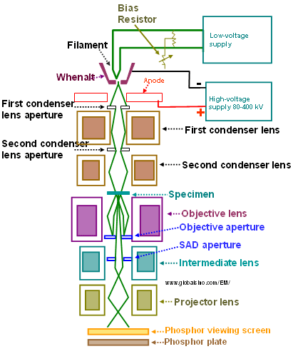
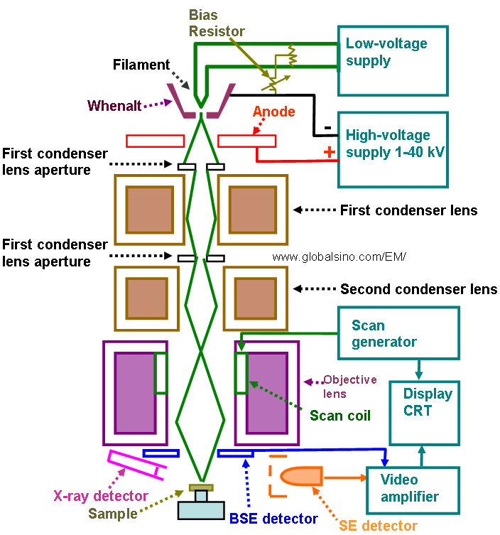
After the seminar, Erin, who is the electron microscopy tech, was kind enough to give Judd, Siwan and I a brief introduction to EM. Electron microscopes work by shining beams of electrons through slides. Areas where the electrons pass through turns up light and where they are scattered turns up dark. Where they passed through the images are much more resolved, creating a usable image that you are able to see. There are two types of electron microscopes present at the Dunn School. There is a transmission electron microscope (TEM) and a Scanning Electron Microscope (SEM).
The TEM sends the electron beam through the sample and creates a flat image that we are able to see through. If you want a 3D image from a TEM, you take a series of images at different angles and compile them on the computer, which is called tomography.
A SEM, however, reads the direction in which the electrons bounce off the surface of the sample, meaning you cannot see through the sample, but it also creates awesome 3D images of what you are looking at. Often times the shocking and interesting images that you see from electron microscopes come from SEM, as they create images that are more aesthetically pleasing. SEM can also be used to find the chemical compositions of different substances based on the x-rays given off by the components, which are read by sensors in the microscope. Both types of electron microscopes start at the top with an electron gun. At the Dunn school, this gun is powered by either a tungsten filament or a small purple crystal. While the crystal creates a stronger, brighter stream of electrons, it is exponentially more expensive. This beam passes through a series of magnetic fields (since electrons are negatively charged they are able to be manipulated using magnets) which
refine the shape of the electron beam. It is then passed through the sample and projected onto the bottom plate, where the scientist can look in. All of this is done in a vacuum as contaminants from the air or even the particles that make up the air would cause the electron beam to be taken off track and possibly cause spots on the image. When using the TEM, specimens must be very thin, well preserved, electron dense, and stable in the vacuum. This is essentially the exact opposite of the natural condition of biological specimens, so preparing disks for the TEM is very difficult.
Small circular grids with tiny mesh are used as the slides for the microscope. When a scientist wants to look at a cell under the microscope, the sample must be embedded in a a small plastic hexagon with a point at the end (it sort of looks like a crystal). The sample is then cut precisely into pieces that are 0.5 to 1 nanometres (nm) thick using a glass or diamond knife.
These are then caught by a "boat" attached to the knife filled with water that prevents the slices from sticking together. This is so thin that vibrations from moving around in the room or even talking can cause the slices to be damaged and can cause damage to the knife. The sample is then placed on the circular mesh and mounted on the TEM sample holder. A sample for the SEM is prepared slightly differently. Circular
metal disks are covered by stickers that hold the sample. The whole circle is then coated in
a fine gold layer about 10nm thick, as this makes the sample conductive. You then place the sample in the SEM and are able to look at the images on the computer.
Time: 17:38
Location: St. Edmund Hall, Oxford, England
This morning began with a lab meeting and a presentation by Helen. She is currently writing and hoping to publish a paper on her work with trypanosomes thus far. She has been working with the role certain proteins play in cell division, specifically BA25 and BA35, which are two of the proteins our very own Bungo discovered. She found that when procyclic form of the cells lack BA25 or BA35, there is no change in their splitting. However, when the bloodstream form lacks these proteins, they turn into 2K1N cells (cells that are not able to duplicate), and eventually die. Therefore, finding a way to remove the BA25 and BA35 proteins from the bloodstream form would provide a cure for the disease they cause. Also, when publishing her paper, Helen would like to provide official names for the BA25 and BA35 proteins (since they were discovered in this lab they do not yet have official names besides Bungo's initials), so if anyone has any good ideas let me know (I already suggested we name them Alexa and it wasn't yet entirely dismissed).

Once in lab, Sam and I began working on preparing an argose gel for DNA separation. The gel takes advantage of the string-like nature of DNA To make the gel, Sam placed two grams of argose in 200mL of buffer solution before adding an indicator that will use electrofourosis to light the DNA up under UV light. Once the gel was poured into trays, Sam added combs that will create wells in the gel. These wells differ in width. The wider the well, the more DNA is able to fit but also the lower the resolution.
 |
| What the gel looks like under UV light |
After lunch there was a seminar by Mitsuhino Yanagida, who is a legendary scientist.
 |
| Mr. Yanagida |
After the seminar, Erin, who is the electron microscopy tech, was kind enough to give Judd, Siwan and I a brief introduction to EM. Electron microscopes work by shining beams of electrons through slides. Areas where the electrons pass through turns up light and where they are scattered turns up dark. Where they passed through the images are much more resolved, creating a usable image that you are able to see. There are two types of electron microscopes present at the Dunn School. There is a transmission electron microscope (TEM) and a Scanning Electron Microscope (SEM).
 |
| SEM |
 |
| TEM |
 |
| TEM image |
 |
| SEM image |
 |
| TEM schematic |
 |
| SEM schematic |
 |
| TEM sample |
 |
| SEM samples |


Comments
Post a Comment