18.6.13
Date: 18.6.13
Time: 11:01
Location: St. Edmund Hall, Oxford, England
This morning in lab began with Jack and a milk rinse of the protein membrane began yesterday. This was to cover the membrane with antibodies so that when the other antibodies were added to the membrane, they would only stick to the proteins, sort of like saturating a solution so that none of a new solid will accidentally dissolve.
 From the lab to the microscope room, the fun continued with Helen. Here we looked at cells that had been treated with a detergent to strip away the membrane and proteins on the outside so that they appeared transparent then tagged with immuno fluorescence to indicate certain parts of the cells. First, we examined a protein that exists right where the cells split, between the two skeletons of the cells. This protein is present in both the human and fly form of the parasite, however the fly form does not require this protein to duplicate. On the contrary, the human form does. This means that by eliminating this protein in the human form, the parasite will not be able to split and will die. Finding a way to eliminate this protein in the fly form of the parasite could bring about a cure to HAT. The second indicator showed the flagella connector, the part where the old and new flagella of a splitting cell convene. However, what I found throughout all these tests is that some trials are more successful than others and that science is about trying things, failing, trying again, failing and trying once more.
During a brief intermission Helen B told me about the process by which the lab goes about acquiring the antibodies used for the tests. Each antibody has a specific portion of the cell that it wants to target. There are antibodies for the
nucleus, flagellum, certain proteins, etc. In order to create these antibodies, foreign agents, whatever the antibody is supposed to detect, are injected into an animal (mice, goats, sheep, etc.) so that that animals immune system creates the appropriate antibodies, that are then collected and bottled for later use. Over the twenty years his lab has been in operation, Professor Gull has acquired quite a collection of antibodies that he generously shares with other labs across the world, free of charge. Helen shared that one large challenge of labs such as this one is that the cost of acquiring materials, such as these antibodies, from large pharmaceutical companies is too great, preventing scientists from doing experiments that could potentially result in breakthroughs.
After lunch, the process of creating new slides began. First, the medium for the cells is created and the cells are added. Using methane to stick the cells to a preprepared microscope slides, the samples are treated with detergent to strip the cells of proteins and membranes so that they become translucent,
 which makes reading the samples under the microscope easier. The samples were then washed with antibodies twice, adding the fluorescent indicator to the antibodies in the second antibody wash.
 The final part of the day consisted some fun with the electron microscope. Cells were treated with an antibody wash, much like what was looked at under the normal microscope, however instead of fluorescent tags, the cells were tagged with gold atoms, 10 nanometers wide. These gold atoms were attaching to proteins in the Flagellum Attachment Zone (FAZ), the area where the flagellum attaches to the cell body. The electron microscope, which shoots rays of electrons through a phosphoric lens to produce a green color, creates images by bouncing the electrons off of objects, empty spaces come back green and light while electrons that come in contact with the cells are scattered, creating a dark area. Electron microscopes are very difficult to work and quite large, so are suitable only for lab environments.
Time: 11:01
Location: St. Edmund Hall, Oxford, England
This morning in lab began with Jack and a milk rinse of the protein membrane began yesterday. This was to cover the membrane with antibodies so that when the other antibodies were added to the membrane, they would only stick to the proteins, sort of like saturating a solution so that none of a new solid will accidentally dissolve.
 From the lab to the microscope room, the fun continued with Helen. Here we looked at cells that had been treated with a detergent to strip away the membrane and proteins on the outside so that they appeared transparent then tagged with immuno fluorescence to indicate certain parts of the cells. First, we examined a protein that exists right where the cells split, between the two skeletons of the cells. This protein is present in both the human and fly form of the parasite, however the fly form does not require this protein to duplicate. On the contrary, the human form does. This means that by eliminating this protein in the human form, the parasite will not be able to split and will die. Finding a way to eliminate this protein in the fly form of the parasite could bring about a cure to HAT. The second indicator showed the flagella connector, the part where the old and new flagella of a splitting cell convene. However, what I found throughout all these tests is that some trials are more successful than others and that science is about trying things, failing, trying again, failing and trying once more.
During a brief intermission Helen B told me about the process by which the lab goes about acquiring the antibodies used for the tests. Each antibody has a specific portion of the cell that it wants to target. There are antibodies for the
 |
| Antibodies with an indicator binding to a cell |
After lunch, the process of creating new slides began. First, the medium for the cells is created and the cells are added. Using methane to stick the cells to a preprepared microscope slides, the samples are treated with detergent to strip the cells of proteins and membranes so that they become translucent,

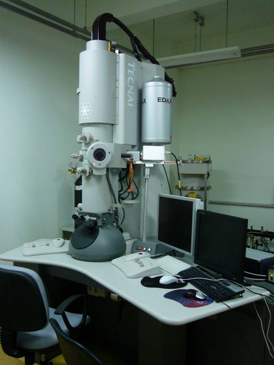 The final part of the day consisted some fun with the electron microscope. Cells were treated with an antibody wash, much like what was looked at under the normal microscope, however instead of fluorescent tags, the cells were tagged with gold atoms, 10 nanometers wide. These gold atoms were attaching to proteins in the Flagellum Attachment Zone (FAZ), the area where the flagellum attaches to the cell body. The electron microscope, which shoots rays of electrons through a phosphoric lens to produce a green color, creates images by bouncing the electrons off of objects, empty spaces come back green and light while electrons that come in contact with the cells are scattered, creating a dark area. Electron microscopes are very difficult to work and quite large, so are suitable only for lab environments.


Comments
Post a Comment