21.6.13
Date: 21.6.13
Time: 14:40
Location: Dunn School of Pathology, Oxford, England
Today was the graduate student symposium, where first, second, and third year students presented the work they have done thus far and work they plan on doing in the future. First and second year students prepare posters explaining their work, while third year students give 15 minute talks with questions.
First up was Narin Hengrung who discussed his work with the structural properties of influenza polymerase. The portion of the influenza virus he is studying is incredibly small and not much is known about its structure. He is studying the PB1, PB2 (which are basic components), and the PA (which is an acidic component). In order to gain more information on the structure he looked at other viruses that have similar characteristics to the influenza virus.
 He also discussed the many problems he encountered during his work, reminding me that you fail often in science and that acknowledging and sharing your mistakes is part of the learning process. The actual appearance of the presentation was nice. Set on a clean white background, there was no text, just colourful graphs and diagrams showing the results of his data. With smooth transitions and plenty of blank space, the presentation was very good. The only negative I would note is that, while the lack of text was refreshing, if you missed a sentence or two of what he was saying, there was no on-screen reference to look to (though that may have just been my difficulty understanding his accent).
The second presentation came from Alison Leishman who talked about Lysosomal Storage Disorder (LSD--trippyy). This disorder comes from a mutation in the human genome that does not allow for proper storage in the lysosome in newborn children. This causes problems when the lysosome ruptures and spills the contents into the patient's body.
 While not a problem when occurring in one or two cells, this rupturing occurs many times over, causing death either right after birth or within the first year of life. In order to combat this disease, Alison is using a vitamin D3 treatment that has so far been successful. She also realized that by treating a particulate that left the lysome with enzymes, the problem was slightly alleviated. Her actual presentation, in contrast with Narin's, contained text that clearly allowed the audience to follow along her thought process. She also included many graphs and diagrams showing her results and "snaked", or made eye contact throughout the room as well as used her hands well while she talked. The only problem with her presentation, which I gleaned from the questions afterwards, was that the enzyme treatment that she is working on might not be as effective as she might hope due to the need to use a different enzyme for each patient and given that time is critical in an illness such as LSD her solution may not be feasible in all cases.
The third talk before the tea break came from our very own Michael Fiebig. His talk about leishmania mexicana, (which he practiced giving earlier in the week), was much improved from his initial effort. I will not go into the details of his presentation because this blog is all about leishmania.
 Though I will comment about his power point. While there was a lot of text that he read directly off his slides, he had good projection and character as well as a bit of humour. His transitions were set up so that each line of text came up with a click of the keyboard which I personally was not a fan of but it allowed the audience to follow along with Mike and not get distracted. Oh, and I also got mentioned in the acknowledgements as a member of the lab!! Woo-hoo!!
After a break for tea and biscuits as well as time to mill around and look at first and second year posters, the presentations resumed.
The first presenter after the break was Olga Kuznetsova, who talked about changes in POI II CTD code during development. During RNA processing, the CTD can change. Olga looked at these changes in human stem cells, which has never been done before. The rest of the presentation contained many protein codes and abbreviations that I did not fully understand. Her entire presentation was divided into two parts, the first part explaining her involvement with CTD and the second with RPAP2. Her power point, done entirely in black and white, contained medium amounts of text as well as graphs. Her results featured graphs of data as well as Westerns.
The next presenter, Chris Duncan, discussed his work with HIV-1 in t-cells and its methods of transmission between cells. The t-cells that contained CD-4 were usually the ones that then became infected with the HIV virus. T-cells, which usually live relatively short lives, are alive much longer when infected. Chris' work focused mainly on the ways in which the HIV virus is transmitted between cells once the patient is infected. HIV, which uses the incredibly efficient cell-to-cell transmission (vs. cell-free transmission) was found to be spread more easily from the macrophages as opposed to other t-cells. This is because the macrophages have pockets where the virus is able to mature before being transmitted, increasing transmission rate and efficiency Chris' power point contained lots of colours as well as some text. He did a great job showing his experimental process through diagrams and pictures and showed his results in an organized fashion. He also included a movie showing the transmission of the virus, tagged with a fluorescent antibody, between a macrophage and t-cell, which was a great visual aid.
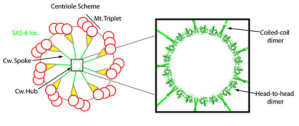
Third was a presentation on the structural form of centriole by Matthew Cottee.
 Matthew is a structural biologist, meaning he studies the structural geometry of living organisms trying to understand why they look the way they do, and what each component within the structure does. Centrioles forms centrosomes, and improper construction of these centrosomes can result in a genetic disease called Microcephaly, where the brain and head are smaller than normal.
 It appears as though this size difference does not cause any developmental problems, as the structure of the brain is exactly the same as a healthy brain, only smaller. Matthew's work has deduced thus far that the number of lose centrosomes is directly proportional to the number of incorrect bonds.
Lunch was only the halfway point of the symposium!
Next was Sofia Nordlander who presented on her work regarding the flagellin sensor NLRC4 protection against a mucosal infection. Her work is focused on Inflammatory Bowel Disease (IBD). In the small intestine, there is bacteria and, on the other side of a wall of epithelial cells, immune cells which must react appropriately when bacteria invades the cell wall. There are also flagellin, which are abundant in the intestinal wall. Sofia performed her tests in mice, in which she manipulated the NLRC4 values of either the epithelium or immune cells, or both. Her results showed that mice with epithelium deficient in NLRC4 acted like mice that were totally NLRC4 deficient. This means that the epithelium needs to contain NLRC4 or the mice will not be able to stop cell colonization or spreading. Sofia's presentation was well laid out, contained a plethora of results, graphs, and diagrams to display her ideas. Her thought process was easy to understand and to top it all off her experiments worked!
We were then given a break for afternoon tea and crumpets (how civilized!) before the presentations resumed.
Himadri Mukhopadyay gave a presentation regarding a systems approach to t-cell receptor proximal signaling. T-cells, which detect infections in the body, are a crucial part of the immune response. Himadri is studying how t-cells communicate with each other and how they communicate within themselves. In order to do this, he created mathematical equations and graphed these equations to show how the t-cells should behave. He used the Hill Function to show the predicted reaction of the t-cells then manipulated the equation to show how the cell would react in certain situations.
Himadri also discovered that when the number of receptor sites on the t-cell is decreased, the potency of the immune response is also decreased. The opposite is also true. Increasing the amount of receptor sites increases the potency of the response. The subject matter that Himadri is studying is incredibly important and he did a good job presenting. However he ran over time by a great deal and wasn't able to finish presenting his findings. Also, I had a question that I did not have the opportunity to ask during the question section which was, wouldn't a mathematical model only account for ideal conditions, which are not present in the real world? (Similar to equations in chemistry which only apply to ideal gases or compounds at ideal conditions (STP), which do not exist in the lab).
Focusing Antibody Responses to Certain Proteins with Site Specific Tags was given by Torben Schiffner. This project mainly discussed HIV and the Influenza virus. These two are similar because they both put out false indicators so that the immune system creates antibodies that go to attack the virus but, much like a matador, the indicator is just a smoke screen that the virus then retracts, leaving the antibodies confused and the virus continuing to spread. With the use of glycan masking, Torben discovered that he could cover these false indicators so that the antibodies would not go there, and then also redirect the antibodies to the actual virus so that they could destroy it. Torben's presentation contained a good combination of text, graphs and results, diagrams, and color, however there were times where it was a bit confusing as to what he was talking about and where the information ended and his personal work began.
Finally, Zsofia Novak closed the symposium with a presentation on Centriole duplication. Centrioles are cell structures found in most eukaryotes.
Improper duplication, leaving too many or too few, causes disease. When the centrioles divide, they divide into two parts, a mother and daughter component. The daughter then becomes a mother as a daughter forms and the mother remains a mother as another daughter forms.
 This process repeats itself, meaning the number of centrioles doubles with each division. Zsofia is looking for an Asl-GFP protein on either the mother or daughter portion of the centriole to see where it is located in the cell structure and where it goes during duplication. She found that, as expected, the mother contained larger amounts of Asl-GFP than the daughter and that with the addition of DSas4, the daughter's acquisition of Asl-GFP could be blocked. Zsofia's power point was white on black which made her numerous pictures, graphs, and diagrams stand out. She did a good job highlighting areas she was talking about or areas where she altered the centriole and also included a few movies of time lapses of the centriole growth, which were great visual aids.
While all these people were reporting their data, I was taking some of my own. Science is not famous for its large female presence, however I was surprised to see the number of women presenting. What I found was that

Time: 14:40
Location: Dunn School of Pathology, Oxford, England
Today was the graduate student symposium, where first, second, and third year students presented the work they have done thus far and work they plan on doing in the future. First and second year students prepare posters explaining their work, while third year students give 15 minute talks with questions.
First up was Narin Hengrung who discussed his work with the structural properties of influenza polymerase. The portion of the influenza virus he is studying is incredibly small and not much is known about its structure. He is studying the PB1, PB2 (which are basic components), and the PA (which is an acidic component). In order to gain more information on the structure he looked at other viruses that have similar characteristics to the influenza virus.

The second presentation came from Alison Leishman who talked about Lysosomal Storage Disorder (LSD--trippyy). This disorder comes from a mutation in the human genome that does not allow for proper storage in the lysosome in newborn children. This causes problems when the lysosome ruptures and spills the contents into the patient's body.

The third talk before the tea break came from our very own Michael Fiebig. His talk about leishmania mexicana, (which he practiced giving earlier in the week), was much improved from his initial effort. I will not go into the details of his presentation because this blog is all about leishmania.

After a break for tea and biscuits as well as time to mill around and look at first and second year posters, the presentations resumed.
The first presenter after the break was Olga Kuznetsova, who talked about changes in POI II CTD code during development. During RNA processing, the CTD can change. Olga looked at these changes in human stem cells, which has never been done before. The rest of the presentation contained many protein codes and abbreviations that I did not fully understand. Her entire presentation was divided into two parts, the first part explaining her involvement with CTD and the second with RPAP2. Her power point, done entirely in black and white, contained medium amounts of text as well as graphs. Her results featured graphs of data as well as Westerns.
The next presenter, Chris Duncan, discussed his work with HIV-1 in t-cells and its methods of transmission between cells. The t-cells that contained CD-4 were usually the ones that then became infected with the HIV virus. T-cells, which usually live relatively short lives, are alive much longer when infected. Chris' work focused mainly on the ways in which the HIV virus is transmitted between cells once the patient is infected. HIV, which uses the incredibly efficient cell-to-cell transmission (vs. cell-free transmission) was found to be spread more easily from the macrophages as opposed to other t-cells. This is because the macrophages have pockets where the virus is able to mature before being transmitted, increasing transmission rate and efficiency Chris' power point contained lots of colours as well as some text. He did a great job showing his experimental process through diagrams and pictures and showed his results in an organized fashion. He also included a movie showing the transmission of the virus, tagged with a fluorescent antibody, between a macrophage and t-cell, which was a great visual aid.
Third was a presentation on the structural form of centriole by Matthew Cottee.

Lunch was only the halfway point of the symposium!
Next was Sofia Nordlander who presented on her work regarding the flagellin sensor NLRC4 protection against a mucosal infection. Her work is focused on Inflammatory Bowel Disease (IBD). In the small intestine, there is bacteria and, on the other side of a wall of epithelial cells, immune cells which must react appropriately when bacteria invades the cell wall. There are also flagellin, which are abundant in the intestinal wall. Sofia performed her tests in mice, in which she manipulated the NLRC4 values of either the epithelium or immune cells, or both. Her results showed that mice with epithelium deficient in NLRC4 acted like mice that were totally NLRC4 deficient. This means that the epithelium needs to contain NLRC4 or the mice will not be able to stop cell colonization or spreading. Sofia's presentation was well laid out, contained a plethora of results, graphs, and diagrams to display her ideas. Her thought process was easy to understand and to top it all off her experiments worked!
We were then given a break for afternoon tea and crumpets (how civilized!) before the presentations resumed.
Himadri Mukhopadyay gave a presentation regarding a systems approach to t-cell receptor proximal signaling. T-cells, which detect infections in the body, are a crucial part of the immune response. Himadri is studying how t-cells communicate with each other and how they communicate within themselves. In order to do this, he created mathematical equations and graphed these equations to show how the t-cells should behave. He used the Hill Function to show the predicted reaction of the t-cells then manipulated the equation to show how the cell would react in certain situations.
 |
| Hill Function |
Focusing Antibody Responses to Certain Proteins with Site Specific Tags was given by Torben Schiffner. This project mainly discussed HIV and the Influenza virus. These two are similar because they both put out false indicators so that the immune system creates antibodies that go to attack the virus but, much like a matador, the indicator is just a smoke screen that the virus then retracts, leaving the antibodies confused and the virus continuing to spread. With the use of glycan masking, Torben discovered that he could cover these false indicators so that the antibodies would not go there, and then also redirect the antibodies to the actual virus so that they could destroy it. Torben's presentation contained a good combination of text, graphs and results, diagrams, and color, however there were times where it was a bit confusing as to what he was talking about and where the information ended and his personal work began.
Finally, Zsofia Novak closed the symposium with a presentation on Centriole duplication. Centrioles are cell structures found in most eukaryotes.
 |
| Centriole splitting, the daughter portion is the horizontal piece while the vertical piece is the mother |

While all these people were reporting their data, I was taking some of my own. Science is not famous for its large female presence, however I was surprised to see the number of women presenting. What I found was that

the number of boys and girls was very similar, which is unusual not only in science but especially in PhD classes.


Comments
Post a Comment