27.6.13
Date: 27.6.13
Time: 18:09
Location: The Dunn School of Pathology, Oxford, England
Starting off my penultimate day in Oxford, Jack and I helped Richard (who is away doing research in Africa) with his ongoing experiment. He is studying transfection efficiency. When cells are transfected and the new DNA is placed in the cells, cells that have not taken up the new DNA are killed with a drug that the new DNA is resistant to. The cells are then placed in 96-well trays so that some grow, leaving samples of cells with only the new DNA. Richard took cells and put them in trays and then created pairs for each tray with a 1:100 dilution.
We looked at each tray and counted the number of wells where the cells were alive and growing. There were many without any growth, however that should be expected as the only times we should have seen any growth was when there were over 50 wells with growth in the undiluted solution. However, there is also potential for there to be no growth in a diluted solution even if there should be. In one of the trays there were 94 wells with growth in them however the diluted solution contained no growth. While this is unlikely, statistics will tell you that there is actually a chance that that would happen.
Next was a continuation of a transfection of cells that Jack started yesterday. We have seen the beginning of this with Helen the other day and this is the continuation. Jack, who concentrates of the flagella attachment zone (FAZ), is trying to determine where the new flagellum grows from. When the cell is splitting and the new flagella is growing, does it grow like hair, from the body of the cell, or does it grow from the end outward, sort of like stacking Legos on a tower.
The media of the transfected cells was removed before they were placed on the slide. Even though this is the procyclic form of trypanosomes, (which usually sticks to the microscope slides easily), FCS (fetal cow serum) in the media prevents the trypanosomes from sticking to the glass. The media is removed and the whole sample is washed in PBS. It is then treated in detergent which strips the membrane from the cells, leaving only the cytoskeleton. Afterwards the whole slide is dunked in BSA, which acts like the milk in the western blot, effectively blocking any other sites where the YFP might tag proteins. The sample is then placed on the microscope slide and treated with formaldehyde (which is used for embalming and is also found in some keratin hair treatments) which preserves the cells. DAPI and YFP are then added. The DAPI tags the nucleus and kinetoplast of the cells so that they can be seen under the microscope. The YFP (Yellow Fluorescent Protein) will target the new proteins in the FAZ, showing where the new flagellum is growing from. Each life cycle of a trypanosome is eight hours, meaning every eight hours the cell will split. Jack will look at his sample after two hours, as this will ensure that the entire flagella on the cells will not grow yellow because the cell will not be completely full grown yet. In between washes, the slides must remain in a container covered in tinfoil with a wet paper towel at the bottom. This is a humidity chamber, which is used to prevent any of the small amount of liquid on the slide from evaporating.
After lunch I set out placing new pipette tips in empty boxes. Once inside, the boxes were taped up with a special tape designed for the autoclave. Autoclaving is when something is heated to an incredibly high heat so as to sterilize. This tape, which is essentially masking tape with yellow bands on it that turn black when heated to incredibly high temperatures, serves as an indicator for when the tips are finished being sterilized as well as sealing the boxes so that no one can tamper with them. We then brought the boxes down to the autoclave room where there are two giant autoclaves which are essentially giant ovens.
After returning from the autoclave room, Jack and I worked on finishing the slides we had prepared earlier in the morning. The slides are charged, which cause certain liquids, such as water, to behave atypically on them. We had to spend some time trying to spread some water across the wells, as the surface tension on the water was so great it would not wet the sample. They were then placed in his humidity chamber to set.
I then moved over to Francois, who was working on redoing the same tests he did yesterday to deflagellate the cells. Today, he used a sucrose bed that was less dense than yesterday in the hope that the cell bodies would sink to the bottom when centrifuged, which they had not completely done yesterday. He also put them through the centrifuge twice to be sure. To be sure he was getting the maximum gravitational increase possible, he also put his samples through the ultracentrifuge. The ultracentrifuge is like a centrifuge but can go much, much faster, causing a greater increase in gravity. Balancing an ultracentrifuge is the most important process because at speeds upwards of 50,000rpm even the slightest difference in mass would be incredibly dangerous. There are stories of ultracentrifuges that were run unbalanced, causing the machine to fly apart and send the samples through four walls before coming to rest. The ultracentrifuge also works in a vacuum to minimize air friction. While this is not a problem for normal centrifuges, at incredibly high speeds there is a sizeable amount of extra energy that would need to be used to overcome air friction. Francois is using the ultracentrifuge at 50,000rpm for one hour, a force of about 170,000g.
Next we observed Felix, who was in the finally stages of a transfection. He was preparing gel for a DNA run. He was creating a 1% argose gel, which contained 0.5g argose powder in 50mL TAE. This was then heated in the microwave and cooled before a few micro-litres of ethidium bromide was added. It is then immediately placed in the dish with the comb in it, creating the wells the samples would be placed in. This is then set for half an hour. Once the gel had set, he added the DNA samples, tagged with a dye to determine relative sizes once the current had been passed through the gel. Then the system was hooked up to 80V of power for half an hour to allow the DNA molecules to travel.
I then wandered over to tissue culture to watch Sam transfect some cells. He is doing work to determine the location of a certain protein within a cell as well as determine the function of that protein by observing how the cell reacts if the protein is removed. He first prepared three containers of medium before taking his samples to the electrocution machine. He used two separate machines because he needed a higher transfection efficiency for one of his samples. A higher transfection efficiency just means that more cells per unit take up the new DNA. Once the cells are electrocuted, they are not very healthy and about 99% of them die, so as soon as they are electrocuted they are put in the media and allowed about a week's time to recover.
Time: 18:09
Location: The Dunn School of Pathology, Oxford, England
 |
| 96 well plate, round bottom |
We looked at each tray and counted the number of wells where the cells were alive and growing. There were many without any growth, however that should be expected as the only times we should have seen any growth was when there were over 50 wells with growth in the undiluted solution. However, there is also potential for there to be no growth in a diluted solution even if there should be. In one of the trays there were 94 wells with growth in them however the diluted solution contained no growth. While this is unlikely, statistics will tell you that there is actually a chance that that would happen.
Next was a continuation of a transfection of cells that Jack started yesterday. We have seen the beginning of this with Helen the other day and this is the continuation. Jack, who concentrates of the flagella attachment zone (FAZ), is trying to determine where the new flagellum grows from. When the cell is splitting and the new flagella is growing, does it grow like hair, from the body of the cell, or does it grow from the end outward, sort of like stacking Legos on a tower.
 |
| New flagellum growth |
 |
| Nuclei are indicated in blue while kinetoplast are labelled green |
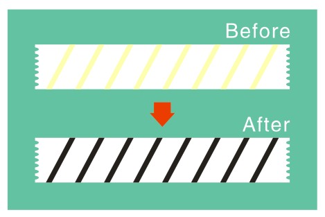 |
| Autoclaving tape |
After returning from the autoclave room, Jack and I worked on finishing the slides we had prepared earlier in the morning. The slides are charged, which cause certain liquids, such as water, to behave atypically on them. We had to spend some time trying to spread some water across the wells, as the surface tension on the water was so great it would not wet the sample. They were then placed in his humidity chamber to set.
I then moved over to Francois, who was working on redoing the same tests he did yesterday to deflagellate the cells. Today, he used a sucrose bed that was less dense than yesterday in the hope that the cell bodies would sink to the bottom when centrifuged, which they had not completely done yesterday. He also put them through the centrifuge twice to be sure. To be sure he was getting the maximum gravitational increase possible, he also put his samples through the ultracentrifuge. The ultracentrifuge is like a centrifuge but can go much, much faster, causing a greater increase in gravity. Balancing an ultracentrifuge is the most important process because at speeds upwards of 50,000rpm even the slightest difference in mass would be incredibly dangerous. There are stories of ultracentrifuges that were run unbalanced, causing the machine to fly apart and send the samples through four walls before coming to rest. The ultracentrifuge also works in a vacuum to minimize air friction. While this is not a problem for normal centrifuges, at incredibly high speeds there is a sizeable amount of extra energy that would need to be used to overcome air friction. Francois is using the ultracentrifuge at 50,000rpm for one hour, a force of about 170,000g.
 |
| This is what happens when you don't balance your centrifuge properly |
I then wandered over to tissue culture to watch Sam transfect some cells. He is doing work to determine the location of a certain protein within a cell as well as determine the function of that protein by observing how the cell reacts if the protein is removed. He first prepared three containers of medium before taking his samples to the electrocution machine. He used two separate machines because he needed a higher transfection efficiency for one of his samples. A higher transfection efficiency just means that more cells per unit take up the new DNA. Once the cells are electrocuted, they are not very healthy and about 99% of them die, so as soon as they are electrocuted they are put in the media and allowed about a week's time to recover.


Comments
Post a Comment